
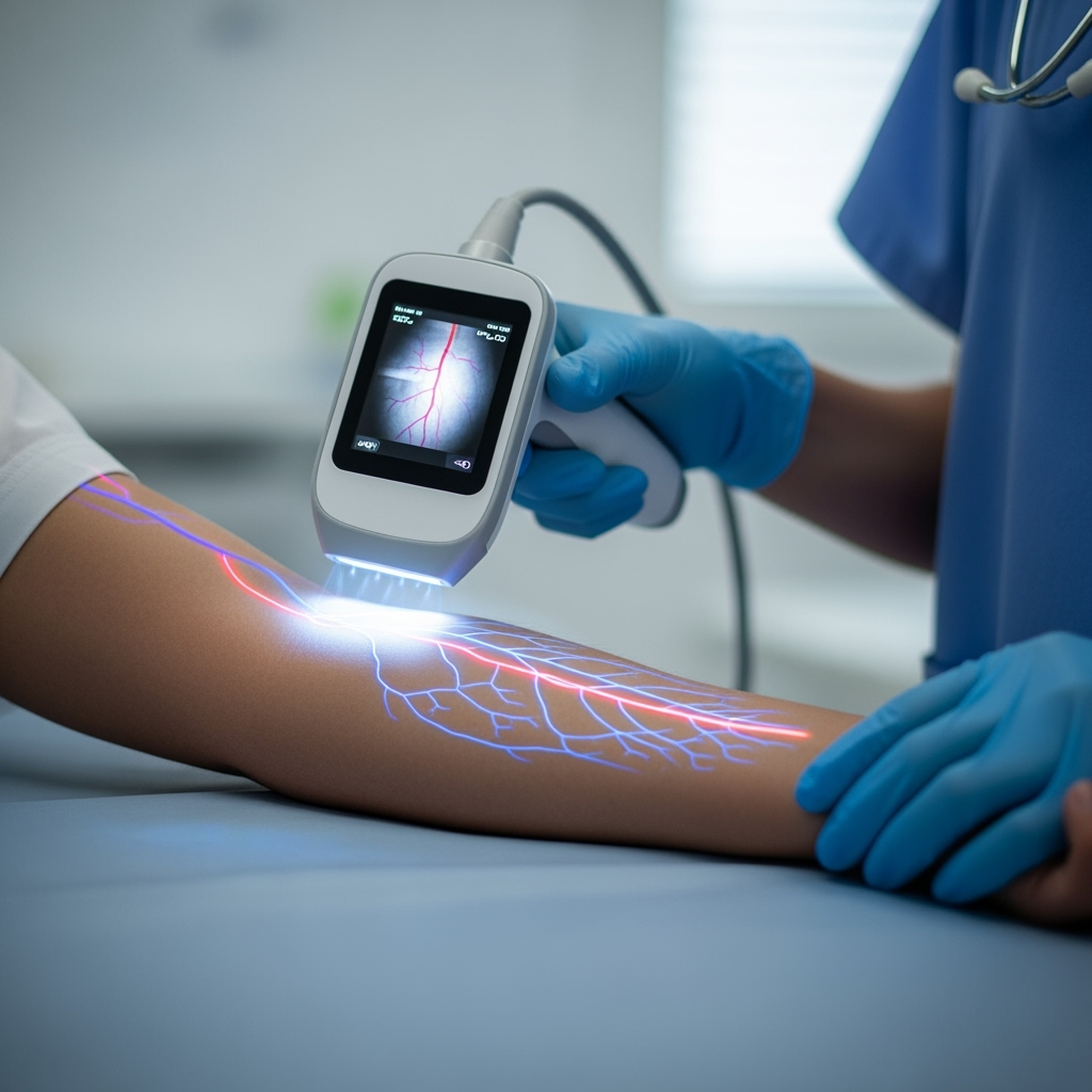
Modern medical diagnostic procedures often involve inserting a needle into a patient’s vein. However, veins may sometimes not be visible, making the procedure difficult for medical staff. This leads to challenges during blood extraction, which can result in errors in laboratory test results. To address this issue, scientists are actively working on developing various visual methods, including near-infrared vein imaging. However, these methods face limitations, such as low depth and poor image contrast. Additionally, the digital image processing algorithms used in these methods are not integrated, with low stability and performance.
Nikita Remezov, a PhD student in the Department of Laser and Biotechnical Systems at Samara University, explained that the algorithm uses efficient operations based on discrete Fourier transforms. He added, ‘Image processing in the Fourier space allows us to effectively enhance areas of the image through sharp changes in intensity between adjacent pixels.’ Remezov noted that ‘the areas within the high-frequency range of the two-dimensional spectrum correspond to sharp transitions in the image. Using the so-called fast Fourier transform, we can use relatively affordable computing units and process images in real time.’
According to Remezov, this new algorithm is part of the development of a local vein imaging device. The device is designed to be cost-effective in terms of manufacturing, efficient, easy to use, and suitable for doctors working with patients who have a high body mass index or other factors that make vein puncture difficult. Researchers are currently working on improving the optical configuration of the future device. By using an image processing algorithm, the device will enable vein imaging in a way that cannot be achieved by other methods, especially in cases of severe skin pigmentation.