
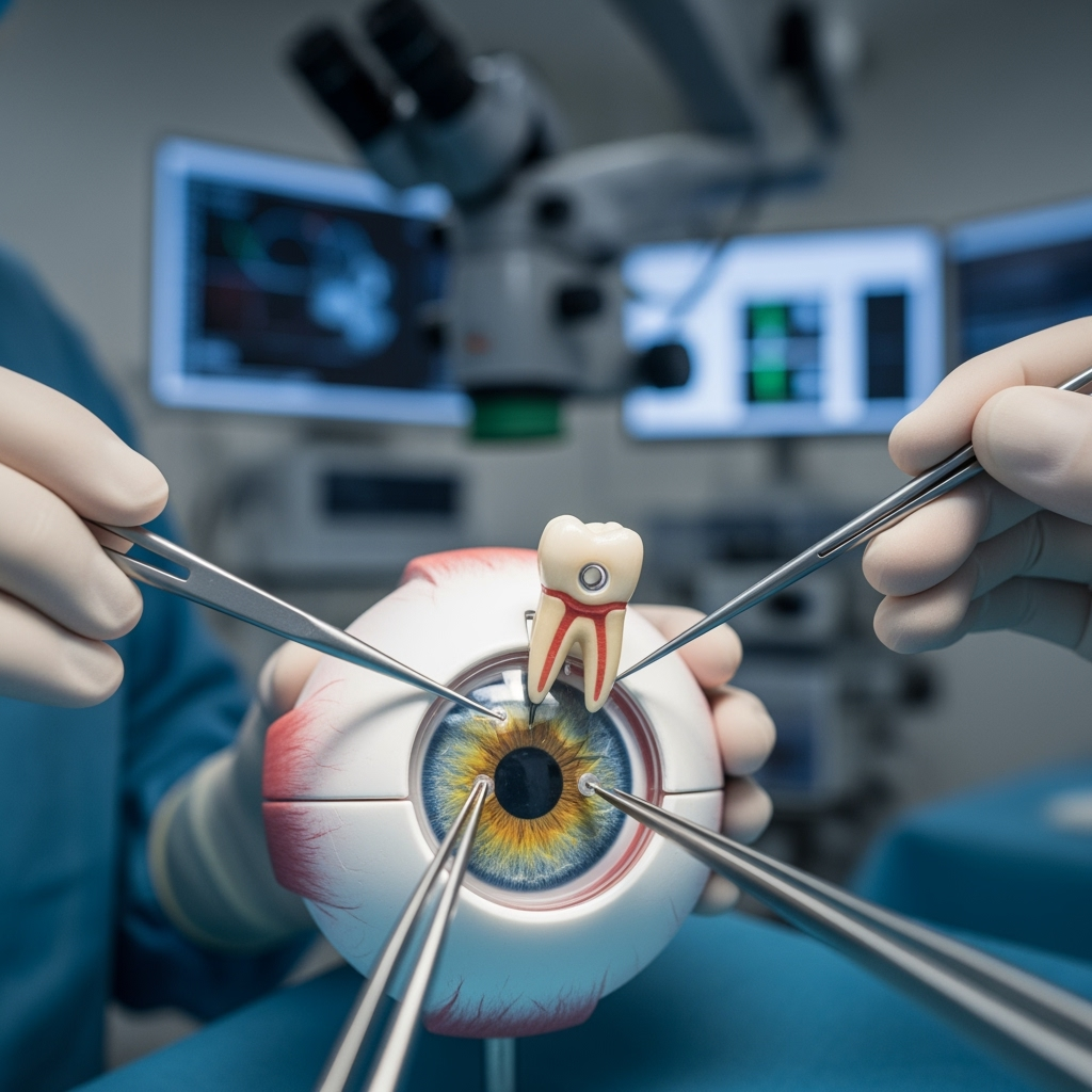
The technique involves taking a portion of a tooth bone and shaping it to hold a plastic cylinder that functions as an artificial lens, as explained by Dr. Ben Kang, Head of the Department of Oral and Maxillofacial Surgery at Vancouver General Hospital in Britain.
Afterwards, the tooth is implanted in the patient’s cheek or eyelid for several months until tissues grow around it. Later, it is fixed at the front of the eye in place of the damaged cornea, according to Western media reports citing the medical team led by Dr. Greg Moloney, Associate Professor of Clinical Corneal Surgery at the University of British Columbia.
Moloney explained that the tooth serves as an ideal structure for securing the lens due to its hardness, durability, and acceptance by the body, as it is part of it. He noted that to create a harmonious appearance, the eye is covered with tissues taken from elsewhere, giving it a distinctive pink color.
Despite undergoing around 50 corneal transplants over two decades, all attempts failed due to his suffering from Stevens-Johnson syndrome, which attacks the stem cells responsible for regenerating the cornea.